
Figure 1.
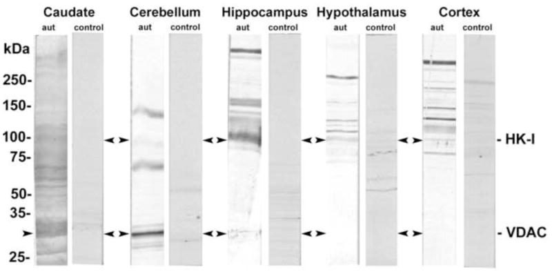
Detection of autoantibodies to normal brain tissue lysates in serum of autistic children. Brain tissue lysates from the caudate nucleus, cerebellum, hippocampus, hypothalamus, and brain cortex were run on 4–20% PAGE and then transferred to nitrocellulose membranes. The membranes were singly incubated with autistic or control sera (Dil. 1:100).