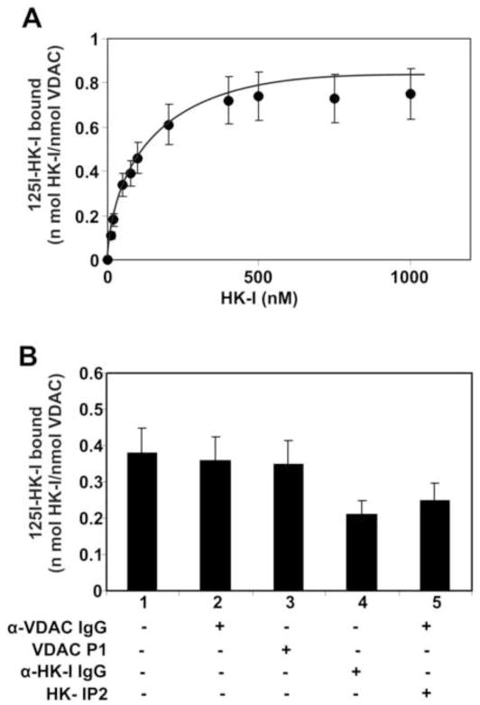
Figure 5.
(A) Binding of HK-I to immobilized recombinant VDAC. Increasing concentrations of [125I]-labeled HK-I were added to VDAC immobilized on 96-strip-well tissue culture plates. The data are represented as mean ± S.D. from experiments performed in triplicate. (B) Inhibition of binding of HK-I to immobilized VDAC. Plates were incubated with a single concentration of [125I]-labeled HK-I (100 nM) and a single concentration (500 nM) of autistic anti-VDAC IgG, VDAC P1, anti-HK-I IgG or HK-I P2. The data are represented as mean ± S.D. from experiments performed in triplicate

