Figure 6.
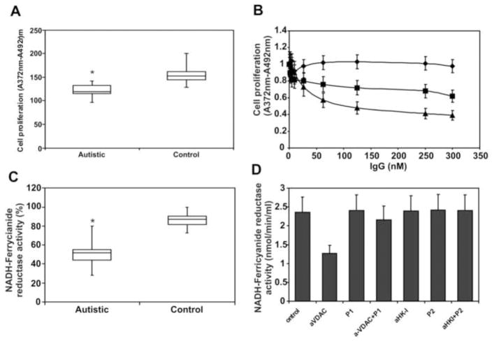
(A) Effect of whole autistic serum (n =34) and isolated autistic autoantibodies on SK-N-SH cell proliferation. Cells in 96-well culture plates (1 × 10⁴ per well) were incubated with 5 μl serum for 48 h in a final volume of 200 μl RPMI 1640 culture medium containing 5% FBS. (B) Increasing amounts of anti-VDAC IgG (▲), anti-HK-I IgG (■), or non-immune control IgG (◆) for 48 h in RPMI 1640 culture medium containing 5% FBS. (C) Effect of whole autistic serum (n=34) on NADH-ferricyanide reductase activity of SK-N-SH cells grown to confluency in 96-well culture plates. (D) NADH-ferricyanide reductase activity of cell-surface VDAC was measured in SK-N-SH cells grown to confluency in 96-well culture plates. Cells were incubated with a single concentration (200 nM) of anti-VDAC, VDAC P1, anti-VDAC + P1, anti-HK-I, HK-I P2, anti-HK-I + P2, or non-immune control IgG in cell culture medium for 2 h prior to the assay. * Significantly different from controls (P<0.001).

