
FIG. 1.
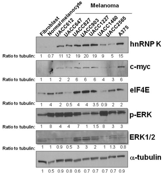
Higher hnRNP K protein level in human melanoma cell lines. Primary fibroblast, primary normal melanocytes, and 8 melanoma cell lines were grown at the log phase and harvested. Western blot analysis was performed using hnRNP K, c-myc, eIF4E, p-ERK, ERK1/2, and α-tubulin antibodies. The intensities of the bands were quantified using ImageJ software and normalized to α-tubulin. The ratio to tubulin was indicated below each band. ERK1/2 was measured as 1 band because of the close distance between these 2 bands