
FIG. 2.
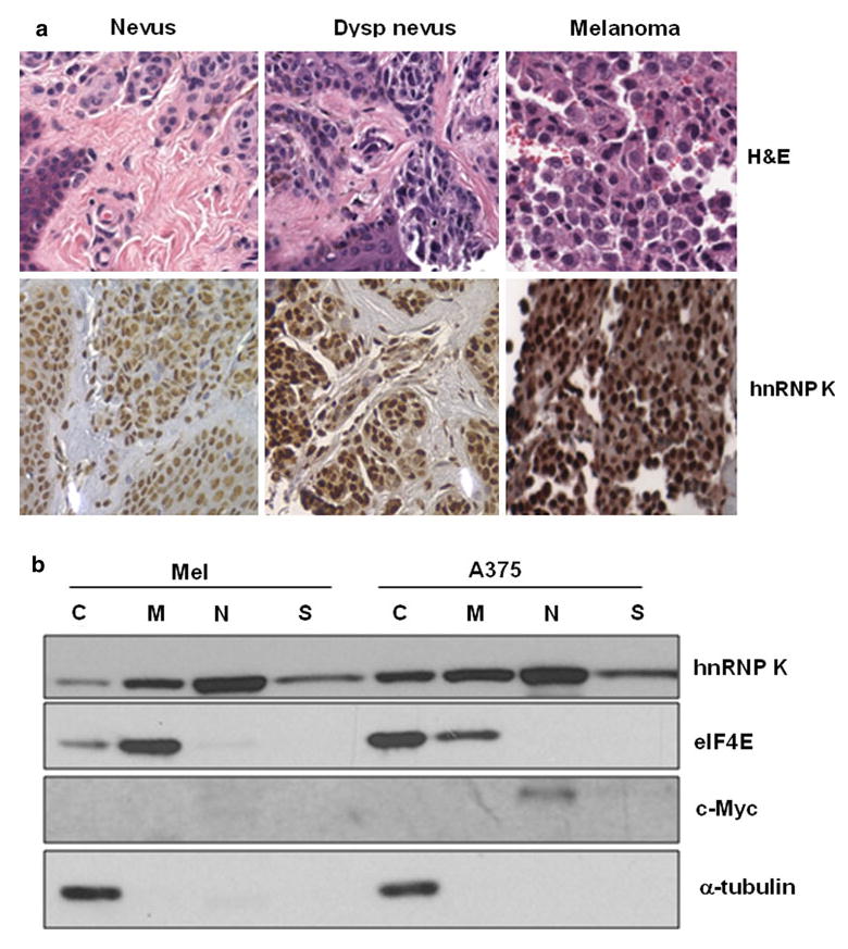
a hnRNP K protein is increased in human dysplastic nevus and melanoma tissues. H&E staining and immunohistochemistry was performed on a melanoma progression tissue microarray using hnRNP K antibody. Representative H&E and immunohistochemistry analysis of the hnRNP K protein in benign nevus, dysplastic nevus, and melanoma tissues were shown. Magnification: 400 ×. b Increased cytoplasmic hnRNP K protein in melanoma cells. Mel-STV (Mel) and A375 cells were fractionated, and Western blot analysis was performed using hnRNP K, eIF4E, c-myc and α-tubulin antibodies. C cytoplasm, M membrane, N nucleus, S cytoskeleton