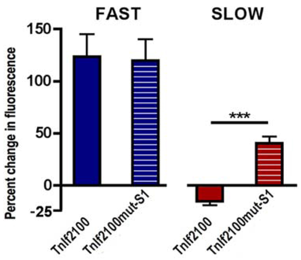
Fig. 2. Mutation of the NFAT site selectively prevents TnIf FIRE repression by slow-patterned activity.
Soleus myofibres transfected by electroporation with TnIf2100 and TnIf2100mut-S1 EGFP reporter constructs and stimulated with either fast or slow activity patterns. To measure enhancer function, myofibres were imaged 1 week after transfection, at the time of denervation and onset of stimulation. A second image was taken after 12 days of stimulation. Bars represent relative changes (%) in EGFP in single soleus fibres stimulated for 12 days with either fast (blue) or slow (red) patterns. Values represent the mean SEM (40 – 72 myofibres from six muscles per condition; ***, P 0.001; one-way ANOVA with Bonferroni post test). This panel is from Rana et al. 2008.

