
Figure 6.
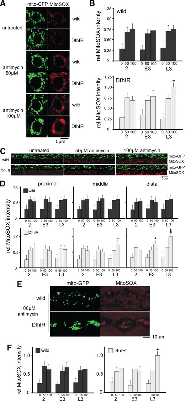
DfhIR neurons do not produce intrinsically higher levels of ROS, but are more susceptible to treatment with the complex III inhibitor antimycin. A–F, Fluorescence intensities of mitoGFP (green) and MitoSOX (red) were used to determine relative levels of ROS production in cell bodies in the ventral ganglion (A, B), axons (C, D), and NMJs (E, F) of DfhIR and wild-type neurons, with and without antimycin treatment, and throughout larval development. Neither wild-type nor DfhIR cell bodies showed increased ROS during development, and DfhIR cell bodies (B, top histogram) did not show higher ROS levels than wild type (B, bottom histogram). Both 50 and 100 μm antimycin treatments elicited higher ROS levels in wild-type and DfhIR cell bodies at all developmental stages, but at late third instar, DfhIR cell bodies responded to antimycin with higher ROS levels than did wild type (B). In axons (C), there were also no significant differences between DfhIR and wild-type ROS levels in any region of the nerve or time in development, but DfhIR axons showed higher ROS levels in response to antimycin in the middle region of the axons at late third instar, and in the distal region at early and late third instar (D). There was also no difference between ROS levels in DfhIR and wild-type NMJs through development, but DfhIR NMJs at late third instar responded to antimycin with higher ROS levels than wild type (E, F). For quantification, all error bars represent the SD and significant differences between DfhIR and wild-type values are indicated (*p < 0.05, **p < 0.01, n = 40 for all experiments).