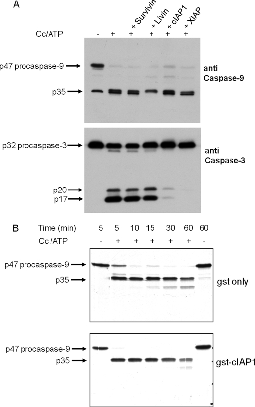
FIGURE 2.
cIAP1 and XIAP prevent the processing of procaspase-3 by the cytochrome c-dependent apoptosome but have no effect on the processing of procaspase-9. A, Western blot analysis of the effects of IAPs on the processing of procaspase-9 and -3. The apoptosome reaction was carried out with S64 from Apaf-1-transfected cells at 1.5 μg/μl for 30 min at 37 °C in the presence or absence of bovine cytochrome c (Cc; 20 μg/ml) plus 1 mm ATP. A sample of the apoptosome reaction (24 μg of protein) was used for SDS-PAGE (12% gel), and Western analysis of caspase-9 and -3 was done as described under “Experimental Procedures.” After immunostaining caspase-9 with the mouse monoclonal antibody, the membrane was reprobed with rabbit monoclonal antibody to cleaved caspase-3. The apoptosome reaction was carried out in the absence of cytochrome c (−) or in the presence of cytochrome c plus the indicated IAP. The concentrations of GST-Survivin, GST-Livin, GST-cIAP1, and GST-XIAP were 1.8, 1.5, 0.27, and 0.13 μm, respectively. B, autoradiogram of the time course of 35S-labeled procaspase-9 processing in the presence of GST (gst only) or GST-cIAP1 (gst-cIAP1). The 20-μl apoptosome reaction included 1.0 μg/μl S64 from Apaf-1-transfected cells and 1 μl of 35S-labeled procaspase-9 and was incubated for the indicated intervals at 37 °C in the presence or absence of bovine cytochrome c (100 μg/ml) plus 1 mm ATP. The reactions contained 500 nm purified GST or 150 nm GST-cIAP1 as indicated. After the indicated intervals, 10 μl of the reaction was fractionated by SDS-PAGE (15% gel) and autoradiographed to visualize 35S-labeled p47 procaspase-9 and the p35 large subunit.

