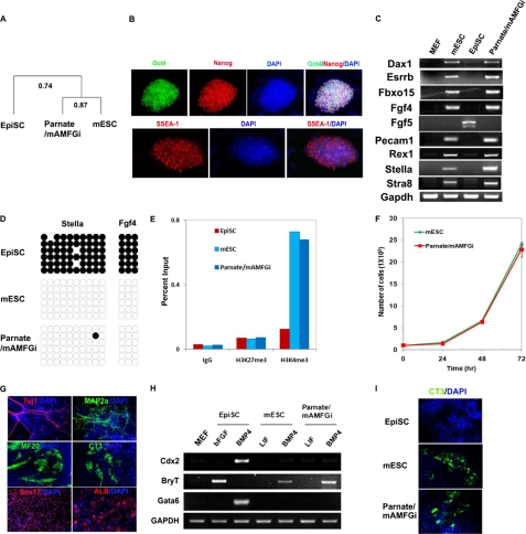
FIGURE 2.
Molecular and functional characterizations of converted Parnate/mAMFGi cells. A, transcriptome analysis of EpiSCs, mESCs, and Parnate/mAMFGi cells showed that Parnate/mAMFGi cells are much more similar to mESCs than to EpiSCs. Two biological replicates were used for all three cell types. B, immunocytochemistry showed homogeneous expression of the pluripotency markers Oct4 (green), Nanog (red), and SSEA1 (red) in Parnate/mAMFGi cells. C, expression of ICM-specific marker genes (Dax1, Esrrb, Fbxo15, Fgf4, Pecam1, and Rex1), germ line competence-associated marker genes (Stella and Stra8), and an epiblast gene (Fgf5) in mESCs, EpiSCs, and Parnate/mAMFGi cells was analyzed by semiquantitative RT-PCR. GAPDH was used as a control. D, the methylation of Stella and Fgf4 promoters was analyzed by bisulfite genomic sequencing. Open and closed circles indicate unmethylated CpG and methylated CpG, respectively. E, the indicated histone modifications in the Stella locus in various cells were analyzed ChIP-qPCR. Genomic DNAs were immunoprecipitated from feeder-free cultured EpiSCs, mESCs, and Parnate/mAMFGi cells with the antibodies as indicated, followed by qPCR analysis using a primer set specific to the endogenous genomic locus encoding Stella. The levels of histone modifications are represented as a percentage of input. IgG served as a no-antibody control. F, Parnate/mAMFGi cells had similar growth rate as mESCs. mESCs and Parnate/mAMFGi cells were passaged every 3 days, and the cell number was counted every 24 h. G, Parnate/mAMFGi cells effectively differentiated in vitro into cells in the three germ layers, including the characteristic neuronal cells (βIII-tubulin- and MAP2ab-positive), cardiomyocytes (cardiac troponin- and MHC-positive), and endoderm cells (Sox17- or albumin-positive). Nuclei were stained with DAPI. H, BMP4 had differential effects on the induction of mesoderm marker (Brachyury), trophoblast marker (Cdx2), and primitive endoderm marker (Gata6) expression in EpiSCs, mESCs, and Parnate/mAMFGi cells. I, directed cardiomyocyte differentiation under monolayer chemically defined conditions demonstrated that Parnate/mAMFGi cells share a similar differentiation response with mESCs and are different from EpiSCs. Cells were characterized with CT3 staining and beating phenotype.

