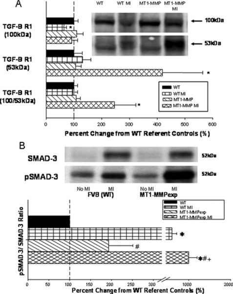
FIGURE 7.
A, LV myocardial membrane extracts from wild type (WT), WT at 14 days post MI, MT1-MMP overexpression (MT1-MMP), and at 14 days post-MI (n = 10/group) were subjected to immunoblotting for transforming growth factor β receptor-1 (TGF-BR1), and the predominant forms were at 53 kDa and a dimer at 100 kDa. The 100-kDa form of TGF-BR1 was reduced in the WT-MI group. TGF-BR1 levels (53 kDa and total) were significantly increased in the MT1-MMP post MI group. B, a common Smad, Smad-2 in the classic TGF signaling pathway, was measured in the same LV myocardial extracts in which both total Smad-2 and the phosphorylated form of Smad-2 (pSmad-2) were quantified. Total phospho-Smad-2 was increased to the greatest degree in the MT1-MMP post-MI group. (*, p < 0.05 versus WT non-MI; +, versus MT1-MMPexp non-MI; #, p < 0.05 versus WT MI.)

