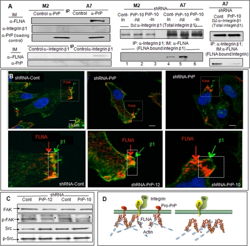
FIGURE 4.
Co-purification of FLNA and integrin β1 and the effects of down-regulation of PrP on integrin β1 and FLNA association. A, left panels, immunoblots (IM) show that PrP co-purifies (IP) with FLNA, and integrin β1 co-purifies with FLNA, but PrP does not co-purify with integrin β1 in A7 cells. This result was confirmed twice. Right panels, immunoblots show that down-regulation of PrP does not alter the expression levels of integrin β1 (top panel) but reduced the amounts of integrin β1 co-purified with FLNA (bottom panel) in the inducible shRNA model (lanes 1–6) and in the constitutively active model. This result was confirmed twice. B, microscopic images show co-localization of FLNA and integrin β1 in control A7 cells. But in PrP down-regulated A7 cells, integrin β1 is separated from FLNA. This result was independently confirmed twice. C, immunoblots show that down-regulation of PrP in A7 cells reduces the level of phosphorylated focal adhesion kinase (p-FAK) but not p-Src. This result was confirmed at least twice. D, a drawing shows that in A7 cells binding of FLNA to pro-PrP pulls FLNA closer to the inner membrane leaflet, which then promotes the binding to FLNA to integrin β1. When the level of PrP is reduced, FLNA retracts from the inner membrane leaflet, which disrupts the binding of FLNA to integrin β1.

