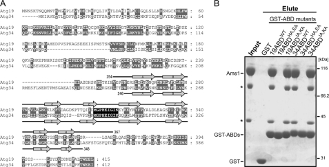
FIGURE 5.
A, sequence alignment between Atg19 and Atg34. Gaps are introduced to maximize the similarity. Conserved or type-conserved residues are shaded gray. Secondary structure elements of the Atg19 and Atg34 ABDs are shown above and below the sequence, respectively. Residues of the DE loop of the ABDs are perfectly conserved (shaded black). B, in vitro pulldown assay between GST-fused Atg19 ABD mutants, GST-fused Atg34 ABD mutants, and Ams1. The input and eluted proteins were subjected to SDS-PAGE and detected by Coomassie Brilliant Blue staining. Asterisks indicate degradation products of Ams1.

