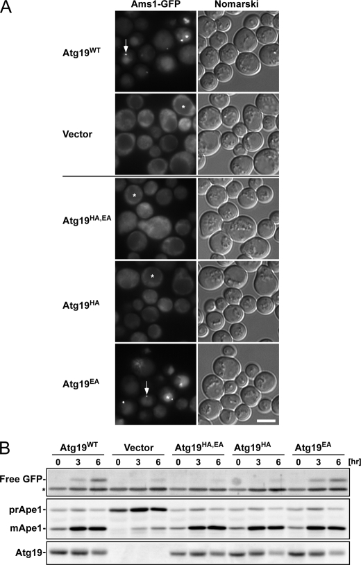
FIGURE 6.
Activity of Atg19 ABD mutants in vivo. A, localization of Ams1-GFP in atg19Δatg34Δ cells expressing Atg19 ABD mutants. Cells were grown in SD + casamino acid medium and then treated with rapamycin for 6 h. Arrows indicate dot structures in the cytoplasm. Asterisks indicate the vacuoles. Scale bar, 5 μm. B, Western blot analyses. Cells used in A were collected at the times indicated after rapamycin addition, and lysates were prepared by the alkaline lysis method. Top, free GFP, produced by the hydrolysis of Ams1-GFP in the vacuole, was detected by immunoblotting using anti-GFP antibody. The asterisk indicates a nonspecific band. Middle, Ape1 maturation was monitored by immunoblotting using anti-Ape1 antibody. Bottom, the accumulation level of Atg19 mutants was detected by immunoblotting using anti-Atg19 antibody.

