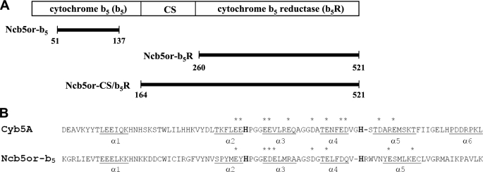
FIGURE 1.
A, schematic diagram of individual domains in human Ncb5or. Cytochrome b5 and cytochrome b5 reductase domains are at the N and C terminus, respectively, with the CS domain in between Ncb5or-b5, Ncb5or-b5R, and Ncb5or-CS/b5R are three constructs used in this study. B, sequence alignment of human Ncb5or (bottom) and human Cyb5A (top) showing all helical segments (i.e. α1–α6 in Cyb5A and α1–α5 in Ncb5or-b5) as well as negatively charged surface residues in core 1 that are conserved in all vertebrate orthologs of each protein (asterisks). In boldface type are heme-ligating residues, His44 and His68 in Cyb5A and His89 and His112 in Ncb5or-b5.

