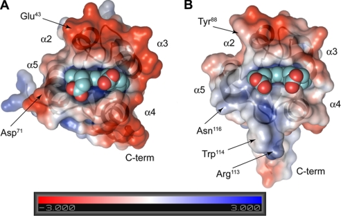
FIGURE 4.
Electrostatic surface maps of bovine Cyb5A (A) and human Ncb5or-b5 (B) show the two proteins in the same orientation as that in Fig. 3A (top). The negatively charged surface in Cyb5A interacting with Cyb5R3 is shown along with a much more weakly charged corresponding surface in Ncb5or-b5. Both proteins are oriented identically with the heme propionates clearly visible.

