
Figure 9.
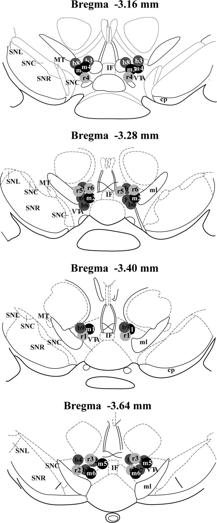
Schematic representation of cannula placements in the VTA. Coordinates of slides are in relation to bregma. b1–b9, NLX mice; m1–m6, chronic morphine + NLX-treated mice; r1–r6, chronic morphine + rp-cAMPS + NLX-treated mice; SNC, substantia nigra compacta; SNR, substantia nigra reticulata; MT, medial terminal nucleus of the accessory optic tract; IF, interfascicular nucleus; ml, medial lemniscus.