
Figure 4. Combination Therapy with anti-CD47 Antibody and Rituximab Eliminates Lymphoma in both Disseminated and Localized Human NHL Xenotransplant Mouse Models.
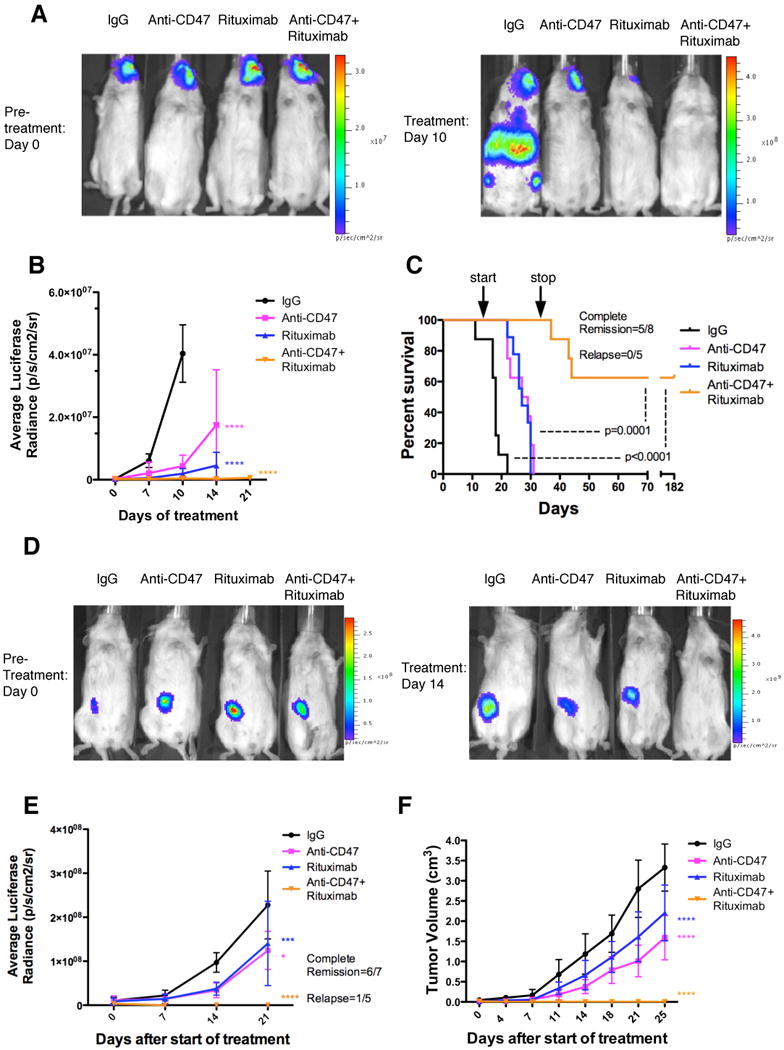
(A) NSG mice transplanted intravenously with luciferase-expressing Raji cells were treated with the indicated antibodies (n=8 per treatment group). Luciferase imaging of representative mice from pre- and 10 days post-treatment are shown (A) and averaged for all mice in each treatment group (B). (C) Kaplan-Meier survival analysis was performed (Table S2). p-values compare IgG control to combination antibody treatment or anti-CD47 antibody/rituximab single antibody to combination. Arrows indicate start (day 14) and stop (day 35) of treatment. (D) Luciferase-expressing Raji cells were transplanted subcutaneously in the flank of NSG mice. When palpable tumors (∼0.1cm3) formed, treatment began with the indicated antibodies. Luciferase imaging of representative mice from each treatment group is shown before (day 0) and during (day 14) treatment. (E) Quantified bioluminescence was determined and averaged for all mice in each treatment group (n=7). (F) Tumor volume was measured with average volume shown. p-values were derived from a two-way ANOVA and compared to IgG treatment. *p<0.05, ***p<0.001, ****p<0.0001. Complete remission was defined as the number of mice with no evidence of tumor at the indicated date. Relapse was defined as the number of mice achieving a complete remission that later developed recurrence of tumor growth. For panel E, one mouse that achieved a complete remission died of non-tumor related causes. Data presented in B, E, and F are mean values +/- SD. See also Figure S4 and Table S2.