
Figure 7. Anti-CD47 Antibody Synergizes with Rituximab Through FcR-Independent and FcR-Dependent Mechanisms.
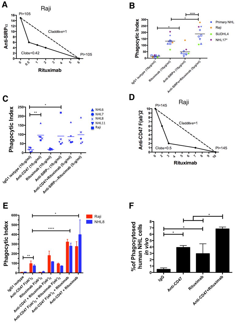
(A) Isobologram analysis of phagocytosis induced by anti-SIRPα antibody and rituximab is shown for Raji cells and mouse macrophages. (B,C) NHL cells were incubated in vitro with the indicated antibodies in the presence of wild type (B) or Fcγr-/- (C) mouse macrophages, and the phagocytic index was determined. (D) Isobologram analysis of phagocytosis induced by anti-CD47 F(ab′)2 antibody and rituximab is shown for Raji cells and mouse macrophages. (E) NHL cells were incubated with wild type mouse macrophages in the presence of the indicated full length or F(ab′)2 antibodies (single antibodies at 10μg/ml, combination antibodies at 5μg/ml each) and the phagocytic index was determined. (F) The level of in vivo phagocytosis measured as the percentage of mouse macrophages containing phagocytosed GFP+ Raji cells (hCD45-GFP+F4/80+) was determined by flow cytometry of livers from mice engrafted with GFP+ Raji cells and then treated with the indicated antibodies (see methods), with each treatment group performed in duplicate. Compared to IgG control, anti-CD47 antibody and rituximab enabled increased levels of phagocytosis. Compared to anti-CD47 antibody alone, combination anti-CD47 antibody and rituximab enabled higher levels of phagocytosis. *p<0.05, **p<0.01, ****p<0.0001. See also Figure S7.