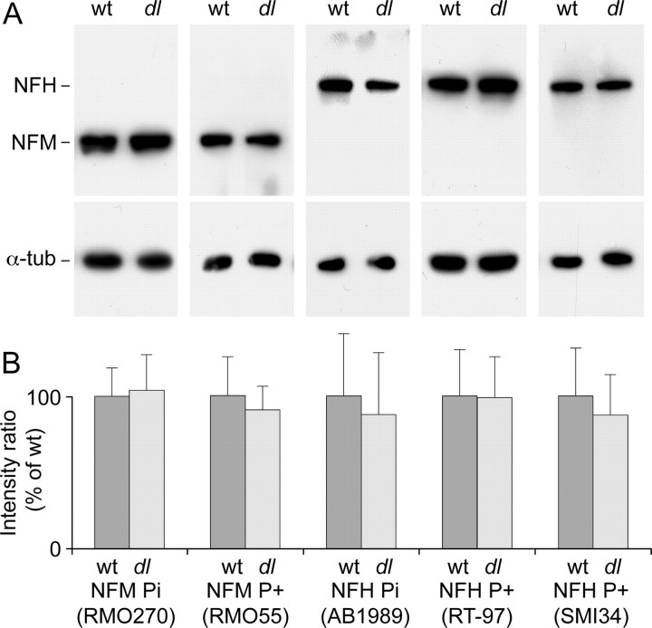
Figure 8.
Neurofilament phosphorylation state in wild-type and dl20J mice. A, Western blots of mouse spinal cord homogenates from wild-type (wt) and dl20J dilute lethal (dl) P4 mice. The blot membranes were cut in half, and the upper half was probed with neurofilament Ab, whereas the lower half was probed with tubulin Ab (α-tub). NFM was detected with mAb RMO270, which binds in a phospho-independent manner. Phosphorylated NFM was detected with mAb RMO55, which binds to phosphorylated epitopes on NFM in a phospho-dependent manner. NFH was detected with pAb AB1989, which binds in a phospho-independent manner. Phosphorylated NFH was detected with mouse mAbs SMI34 and RT97, which bind to phosphorylated epitopes on NFH in a phospho-dependent manner. Tubulin served as a loading control and was detected with mAb B-5-1-2. B, Quantification of blot staining intensities. Each blot was performed in triplicate. For each lane on each blot, the background-corrected intensity of the neurofilament band was divided by the background-corrected intensity of the corresponding tubulin band. For each neurofilament Ab, the resulting intensity ratios were then averaged and normalized to the average for the wild type. The error bars represent the SD about the mean. We observed no significant difference in the intensities of the bands in wild-type and dilute lethal tissue (p = 0.95 for RMO270; p = 0.67 for RMO55; p = 0.73 for AB1989; p = 0.96 for RT97; p = 0.57 for SMI34; Student's t test).

