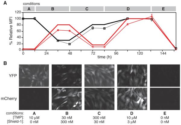
Figure 2. FKBP- and ecDHFR-derived DDs are tunably and predictably regulated using their respective ligands.
(A) NIH3T3 cells doubly transduced with ecDHFR-derived R12Y/Y100I-YFP and the FKBP-derived L106P-mCherry were dosed with varying concentrations of TMP (black) and Shield-1 (red), and fluorescence was monitored by flow cytometry at the time points indicated. Predicted MFI values (thick lines) were generated using a previous dose-response study of the same cell line. Data are presented as the average MFI ± s.d. and normalized against the average MFI of cells dosed with 10 μM TMP and 3 μM Shield-1, which were set at 100%. (B) NIH3T3 cells described in panel A were plated onto 2-well chamber slides and treated for 24 h with concentrations of TMP and Shield-1 corresponding to the conditions used in panel B. Live cells were visualized by epifluorescence microscopy.

