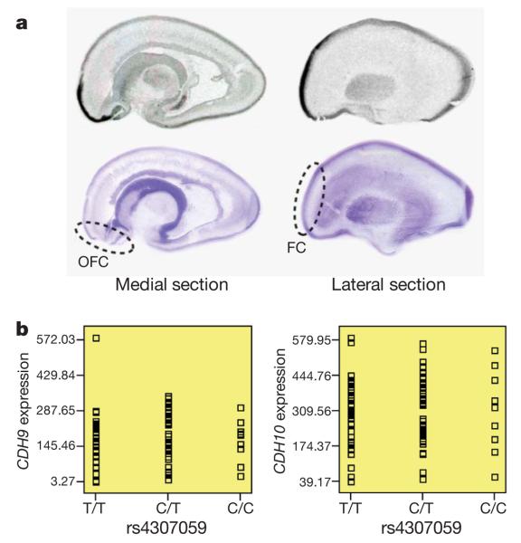
Figure 2. Examination of brain expression for CDH10 and CDH9.
a, The in situ hybridization of CDH10 in human fetal brain sectioned in the sagittal plane. Medial and lateral sections from a representative sample are shown above corresponding cresyl-violet-stained marker slides. Orbitofrontal cortex (OFC) and frontal cortex (FC) are highlighted, with marked expression enrichment. b, The SNP genotypes of rs4307059 are not associated with CDH9 or CDH10 transcript levels in 93 cortical brain tissues.

