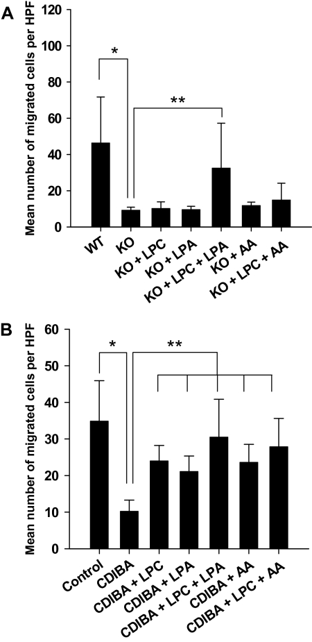
Figure 2.
Effects of lysophospholipids on invasion and migration of cPLA2-deficient and CDIBA-treated vascular endothelial cells. cPLA2α+/+ (wild type [WT]) and cPLA2α−/− (knockout [KO]) murine pulmonary microvascular endothelial cells (A) or murine vascular endothelial 3B-11 cells (B) were added to the top chambers of 24-well transwell Boyden chamber plates containing 8-μm Matrigel-coated inserts. Fresh medium was added to the bottom chambers; vehicle (control) or 2 μM CDIBA (3B-11 only) was added to the medium in both chambers with or without 10 μM lysophosphatidylcholine (LPC), 10 μM lysophosphatidic acid (LPA), or 10 μM arachidonic acid (AA); and the plates were incubated for 24 hours. The inserts were removed and migrated cells on the lower surfaces of the filters were stained with DAPI (4′,6-diamidino-2-phenylindole) and counted in four randomly chosen high-power microscopic fields (HPFs) per sample. Bar graphs plot the mean number of migrated cells per HPF for triplicate samples from three independent experiments; error bars correspond to 95% confidence intervals. A): *P = .004 (Student t test); **P < .001 (analysis of variance [ANOVA]). B): *P < .001 (Student t test); **P < .001 (ANOVA). All P values are two-sided.

