Figure 4.
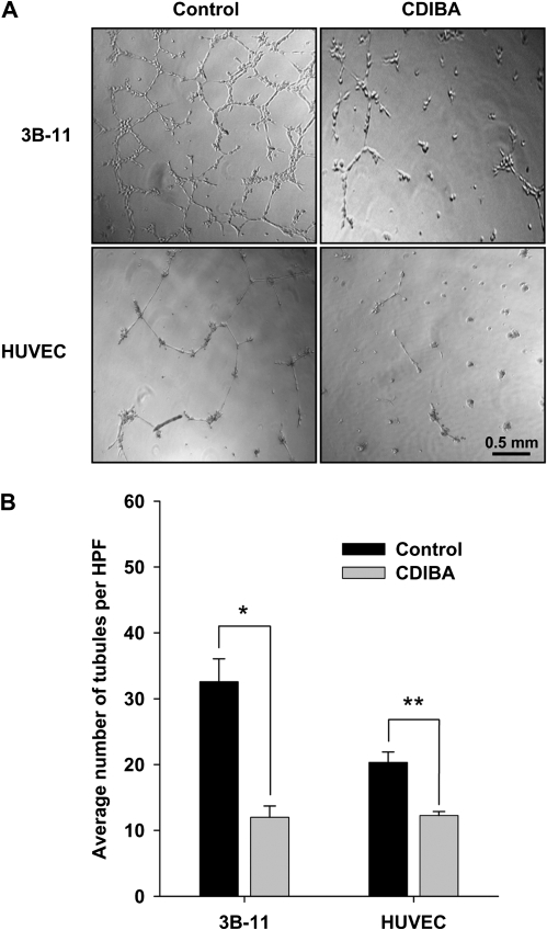
Effect of cPLA2 inhibition on tubule formation by vascular endothelial cells. 3B-11 cells or human umbilical vein endothelial cells (HUVEC) were cultured on Matrigel-coated 24-well plates in the absence (control) or presence of 2 μM CDIBA. Capillary tubule formation was evaluated after 6 (3B-11) or 24 (HUVEC) hours of incubation. Tubule formation was quantified as the number of tubules per high-power microscopic field (HPF) in four randomly selected HPF per sample. A) Representative micrographs of treated cells (at ×20 magnification). B) Bar graph of the mean number of tubules per HPF for 3B-11 and HUVEC from three independent experiments; error bars correspond to 95% confidence intervals. Control vs CDIBA: *P = .005 and **P = .009 (two-sided Student t test).

