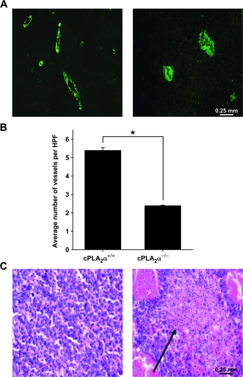
Figure 7.
Vascularity and necrosis in tumors from cPLA2α−/− mice. Lewis lung carcinoma (LLC) cells were injected subcutaneously into the hind limbs of cPLA2α+/+ and cPLA2α−/− mice (n = 6–7 mice per group). Once the average tumor volume reached approximately 700 mm3, the mice were killed by cervical dislocation and their tumors were resected and fixed in 10% formalin. Fixed tumors were then sectioned, and the sections were stained with an antibody against von Willebrand factor (vWF) (an endothelial cell marker) or hematoxylin–eosin. vWF-positive vessels were counted with the use of immunofluorescence microscopy. A) Representative micrographs of positive anti-vWF staining (green) at ×40 magnification in sections of tumors from cPLA2α+/+ (left) and cPLA2α−/− (right) mice. B) Mean number of tumor blood vessels per high-power microscopic field (HPF) from cPLA2α+/+ and cPLA2α−/− mice (three mice per group; six HPFs per slide). Error bars correspond to 95% confidence intervals. *P < .001 (two-sided Student t test). C) Representative micrographs of hematoxylin–eosin-stained sections of LLC tumors from cPLA2α+/+ (left) and cPLA2α−/− (right) mice (at ×40 magnification). Black arrow indicates necrosis.

