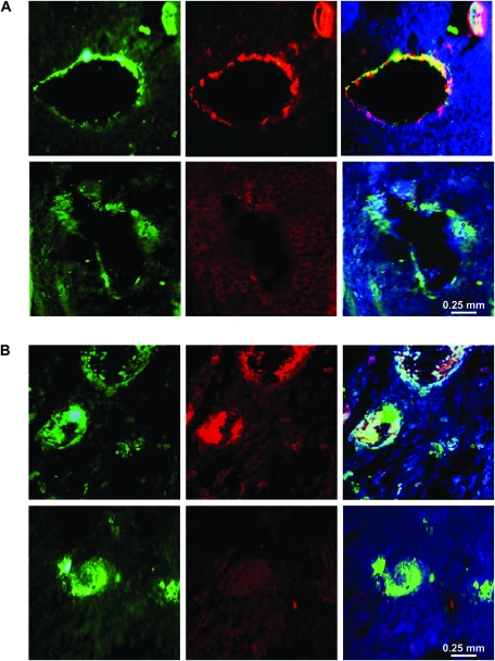
Figure 8.
Pericyte coverage of blood vessels in tumors from cPLA2α+/+ and cPLA2α−/− mice. Formalin-fixed Lewis lung carcinoma (LLC) tumors from cPLA2α+/+ (upper rows) and cPLA2α−/− (lower rows) mice were sectioned and co-stained with antibodies against von Willebrand factor (left panels) and either α-smooth muscle actin (middle panels, A) or desmin (middle panels, B) and counterstained with DAPI (4′,6-diamidino-2-phenylindole). A) Representative micrographs of immunofluorescence staining for von Willebrand factor (green), α-smooth muscle actin (red), and DAPI (blue) in tumors from cPLA2α+/+ and cPLA2α−/− mice (at ×40 magnification). Right panels present merged immunofluorescence staining of von Willebrand factor and cells positive for α–smooth muscle actin (yellow). B) Representative micrographs of immunofluorescence staining for von Willebrand factor (green), desmin (red), and DAPI (blue) in tumors from cPLA2α+/+ and cPLA2α−/− mice (at ×40 magnification). Right panels present merged immunofluorescence staining of von Willebrand factor and cells positive for desmin (yellow).

