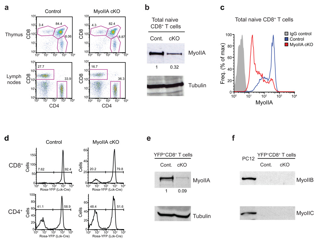
Figure 1. Depletion of MyoIIA in T cells from MyoIIAflox/flox-Lck-Cre conditional knock-out mice.
a) Representative FACS quantification of thymocyte development and peripheral naïve T cells from MyoIIAwt/wt (control) and MyoIIAflox/flox (MyoIIA cKO) mice expressing Lck-Cre. Three pairs of 5–9 week old age-matched mice were used for analysis of thymic development and 6 pairs of mice were used for peripheral T cell quantification. b) MyoIIA expression in peripheral naïve CD8+ T cells from control and MyoIIA cKO mice. Expression was quantified by normalizing to tubulin. c) Control and MyoIIA cKO naïve CD8+ T cells were fixed, permeabilized and stained for MyoIIA expression and analyzed by FACS. d) Frequency of Cre expression in mature single-positive CD8+ and CD4+ T cells in the lymph node determined using a Gt(Rosa26)Sor promoter-loxP-STOP-loxP-YFP (Rosa-YFP) transgenic reporter. e) MyoIIA expression in sorted YFP+-CD8+ peripheral T cells from MyoIIAwt/wt/Lck-Cre+/Rosa-YFP (control) and MyoIIAflox/flox/Lck-Cre+/Rosa-YFP (cKO) mice. Expression was quantified by normalizing to tubulin. f) Lack of Myosin-IIB and Myosin-IIC expression in sorted YFP+-CD8+ control and cKO peripheral T cells. The PC12 cell line was used as a positive control. All data are representative of three independent experiments.

