Figure 1.
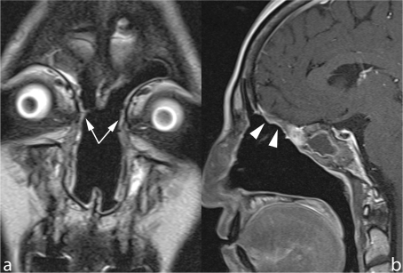
Microendoscopic ethmoidectomy and frontal sinusotomy (Draf-III approach). Coronal TSE T2 (a) and sagittal contrast enhanced SE T1 (b) show the wide communication created between frontal sinuses and nasal fossa (arrows). The duraplasty (arrowheads) exhibits a regular profile, the sphenoid sinus is occupied by thickened mucosa and retained secretions.

