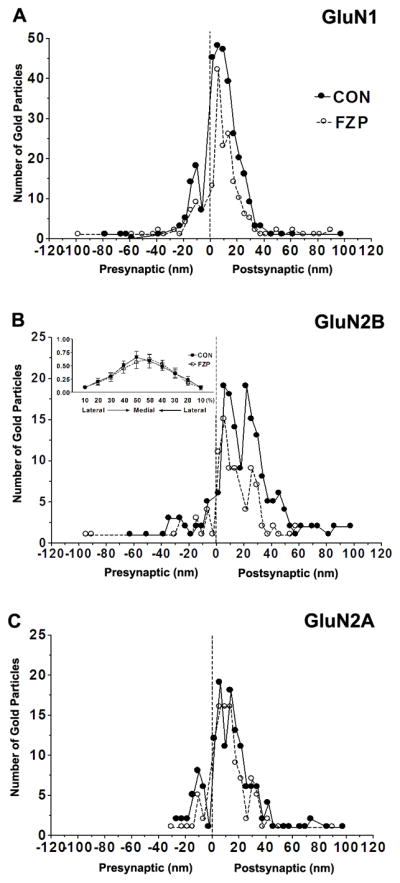
Figure 5. Spatial distribution of GluN1, GluN2A and GluN2B immunogold particles.

(A–C) Distribution of immunogold particles at different distances within the PSD measured in an orthogonal axis from the center of each 10 nm gold particle to the outer leaflet of postsynaptic membrane (up to 100 nm distance) and grouped into 4 nm wide bins (0 on the abscissa represents external face of postsynaptic membrane). Negative values indicate gold particles located in the direction of the presynaptic bouton and synaptic cleft (average width ~20 nm). Positive values indicate immunogold labeling towards the cytoplasm. (A) GluN1 labeling showed one peak, ~6 nm inside the postsynaptic membrane (image resolution ± 2 nm) in control and FZP-withdrawn tissues. (B) GluN2B immunogold in control synapses was similarly distributed in two peaks located 6 and 22 nm within the PSD, but in FZP-withdrawn rats showed a single peak at 6 nm with the peak at 22 nm much reduced. (B, Inset) Lateral distribution profile of GluN2B subunit immunogold labeling. The distance from the lateral edge of the PSD to the center of GluN2B immunogold particles, parallel to the surface of the postsynaptic membrane was measured. The 0–30 nm PSD bin was subdivided laterally into 10 bins consisting of the lateral 10% to medial 50% from either edge of the PSD. GluN2B subunit distribution was concentrated in the middle of the synapse and was not significantly different between control and FZP-withdrawn rats. (C) The spatial distribution of GluN2A labeling in the PSD did not result in clearly separated peaks within the 40 nm width of the PSD and this pattern was unaltered in FZP-withdrawn rats. Data was obtained from 59, 55 and 56 synapses for GluN1, GluN2A and GluN2B-labeled tissues respectively from controls and 66, 51 and 49 synapses from FZP-withdrawn synapses. In all cases, the labeling was primarily found within the 40 nm width of the PSD and the 20 nm surrounding error zone (spatial resolution of indirect immunogold labeling (Matsubara et al., 1996).
