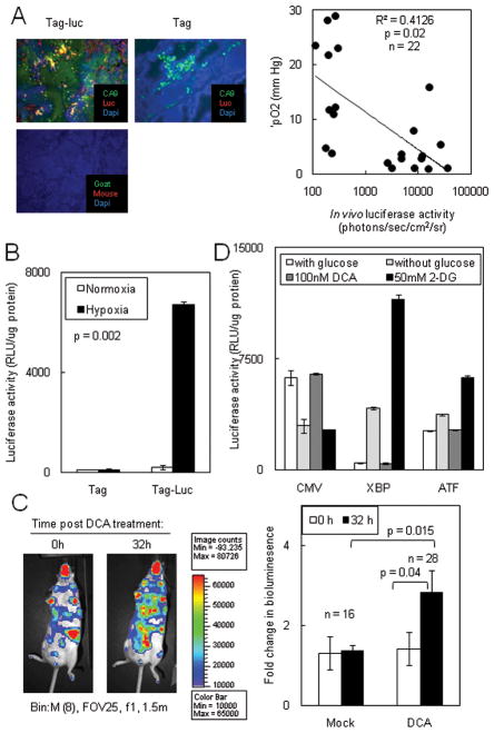
Figure 5. Hypoxia increased XBP1-luc activity.
(A) Left panel: Luciferase co-localized with the hypoxic marker CA-IX. Frozen sections from Tag-Luc and Tag mice were stained with anti-luciferase and anti-CA-IX antibodies and localization of antibodies were detected. Right panel: XBP1-luc activity was plotted against the corresponding pO2 of each tumor. (B) XBP1-luc activity in spontaneous tumors increased under hypoxia. Primary Tag-Luc tumors were cultured in vitro, subjected to normoxia or hypoxia for 48h and luciferase activity was assessed. (C) Left panel: Chemical exacerbation of hypoxia increased ER stress in spontaneous tumors. Tag-Luc mice were treated with vehicle or 50 mg/kg DCA b.i.d. for 2 days and bioluminescence was assessed after 32h. Right panel: Quantitation of bioluminescence in C. (D) DCA alone did not induce ER stress in HT1080 XBP1-luc or ATF4-luc cells.

