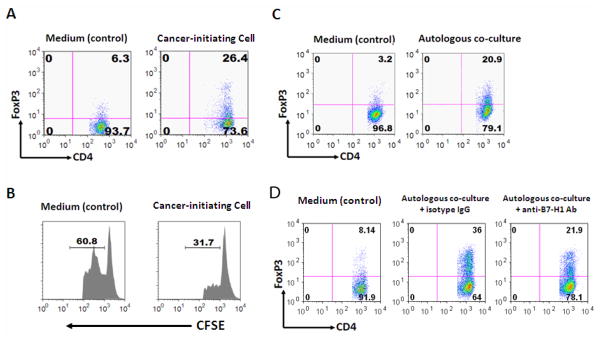
Figure 3. Glioma associated cancer-initiating cells induce functional regulatory T cells.
A. The supernatants from the glioma associated cancer-initiating cells induce an increase of the number of FoxP3+ regulatory T cells in the gated CD4+ T cells as shown by the representative FACS analysis. B. FoxP3+ regulatory T cells induced by the supernatants of cancer-initiating cells suppress T cell proliferation. T cells that were treated with cancer-initiating cell supernatants were harvested, co-cultured for 3 days with autologous PBMCs (labeled with CFSE, responder cells) at a 1:1 ratio in the presence of soluble anti-CD3 and subsequently analyzed via FACScan. The number above the line in each histogram represents proliferating responder cells C. Glioma associated cancer-initiating cells also induce FoxP3+ regulatory T cells through cell-to-cell contact. FACS analysis shows that the glioma associated cancer-initiating cells increased the percentage of FoxP3+ regulatory T cells in the gated CD4+ T cells. D. The induction of Foxp3+ regulatory T cells mediated by cell-to-cell contact is partially reduced after the addition of B7-H1 neutralizing antibody. One representative FACS plot is shown with percentage in upper-right quadrant indicating percentage of positive cells. Similar results were obtained for glioma associated cancer-initiating cells and autologous PBMCs from two other patients.

