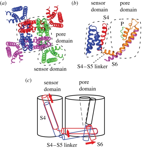
Figure 5.
Structure and conformational change of the potassium channel. (a) Crystal structure of Kv1.2 potassium channel (PDB accession number: 2A79). Each subunit is in a different colour. The pore domain is surrounded by four sensor domains. (b) Structure of a single subunit showing the position of S4 (red), selectivity filter (P), and gate-forming S6 (magenta). (c) Schematic representation of the conformational change that occurs on gating. Blue and red represent the position in the resting and activated state, respectively.

