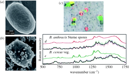
Figure 6.
Bacillus anthracis Sterne spore (a) without and (b) with silver nanoparticle coating. (c) SERS-processed RGB image of a complex bacterial mixture overlaid on BFI. (d) Comparison of extracted Raman spectra from single spores (thin coloured lines), average spectrum (black line) and a library spectrum of BASP (thick red line); (e) extracted Raman spectra from single cells (thin coloured lines), average spectrum (black line) and a library spectrum of BCVG (thick green line). Adapted from Guicheteau et al. (in press). Copyright © Wiley Interscience (2010).

