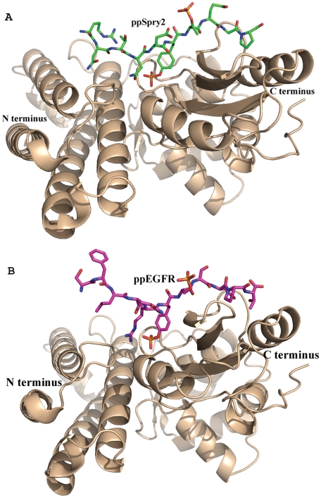
Figure 2. Crystal structure of TKB:ppSpry2 and TKB:ppEGFR complexes.
(A) Ribbon diagram of the TKB:ppSpry2 and (B) TKB:ppEGFR, N- and C- termini are labeled. c-Cbl-TKB is in gold. ppSpry2 (green) and ppEGFR (magenta) peptides are shown in stick represenations. These figures and the following figures in this manuscript were prepared using the program PyMol [40].

