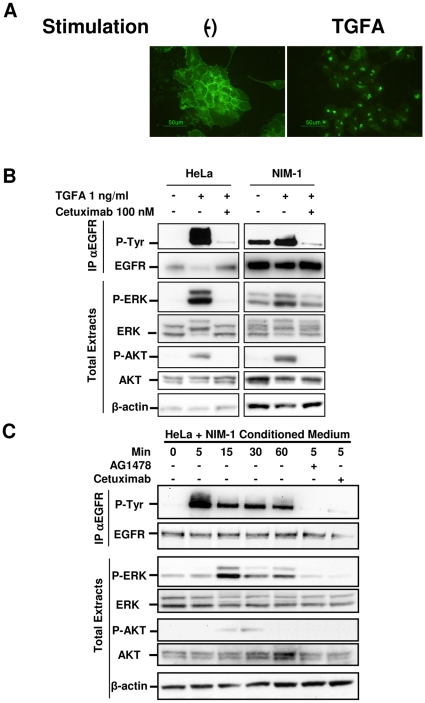
Figure 4. EGFR and TGFA are functional in NIM-1 thyroid cancer cell line.
A. IF analysis with anti-EGFR Ab on serum-starved NIM-1 cells left untreated or treated with 100 ng/ml TGFA for 24 h. B. Biochemical analysis on HeLa and NIM-1 cell extracts. After serum starvation for 24 h, the cells were left untreated or treated with 100 nM Cetuximab, or control chimeric Ab (data not shown) for 2 h, and then stimulated with 1 ng/ml TGFA for 5 min. Abs used are indicated. EGFR phosphorylation has been analyzed by immunoprecipitation (IP) with anti-EGFR Ab and western blotting with anti-phosphotyrosine Ab (P-Tyr). β-actin is shown as a control for protein loading. C. Western blot analysis on HeLa cell extracts. After serum starvation for 24 h, the cells were exposed to fresh medium without FBS (-) or to conditioned medium of NIM-1 cells for 1 h. Pretreatment with AG1478, Cetuximab, or control chimeric Ab (data not shown) was performed for 2 h, and then NIM-1 conditioned medium was added to the culture. As control of specific Ab inhibition, the cells were treated with an unrelated human Ab. Abs used are indicated. β-actin is shown as a control for protein loading. One representative experiment of three is shown.

