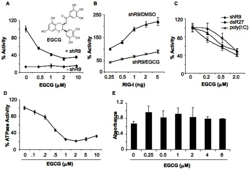
Figure 1. EGCG inhibits RIG-I signaling.
(A) Effect of EGCG titration on triphosphorylated ssRNA, shR9-dependent RIG-I signaling. EGCG was added to HEK293T cells expressing full length RIG-I, IFN-β luciferase and Renilla luciferase reporters as described in the methods. The presence (+ shR9) and absence (- shR9) of shRNA shows the background and induced level of reporter expression in the presence of RIG-I. The activation of signaling obtained upon transfection of shR9 in the absence of EGCG was set as 100%. The chemical structure of EGCG is shown in the inset. The data are shown as a mean +/− standard deviation. (B) Effect of RIG-I plasmid concentration on EGCG inhibition. HEK293T cells were transfected with increasing amounts of pUNORIG-I plasmid that expresses full length RIG-I without changing the amount of reporter plasmids. The total amount of plasmids was kept constant by addition of pUNO vector plasmid. EGCG was added at 2 µM and DMSO was used as control. The data are represented as a mean +/− standard deviation. (C) Activation of RIG-I signaling with single and double stranded RNA agonists was inhibited by EGCG in the reporter assay in HEK293T cells. The data are shown as a mean +/− standard deviation. (D) EGCG inhibits ATPase activity of recombinant full length RIG-I. Different amounts of EGCG were added along with shR9 agonist to the ATPase reaction. ATPase activity obtained with DMSO was considered as 100%. (E) WST-1 assay to determine the toxicity of EGCG in HEK293T cells. The data are shown as a mean +/− standard deviation.

