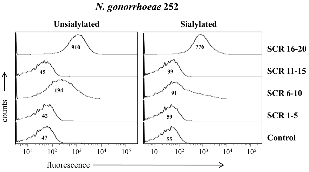
Figure 4. Binding of human fH/murine Fc fusion proteins to N. gonorrhoeae.
Binding of fH derived SCR regions/mouse Fc fusion proteins to unsialylated and sialylated N. gonorrhoeae strain 252. SCR regions spanned the entire length of the fH molecule. Unsialylated strain 252 bound SCR 6–10 and SCR 16–20, sialyated strain 252 bound (only) SCR16–20. Each Fc fusion protein was added at a concentration of 80 nM. The “Control” lacks fusion protein(s). Detection was performed as described in Figure 3. The x-axis represents fluorescence on a log10 scale, the y-axis the number of events. Numbers in the histogram represent the median fluorescence of the bound Fc fusion proteins to N. gonorrhoeae strain 252. One representative experiment is shown of three independently performed experiments.

