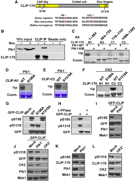
Figure 1.
CLIP-170 is a Plk1/CK2 substrate in vitro and in vivo. (A) Schematic of CLIP-170 with two-identified Plk1/CK2 phosphorylation sites. * marks the two phosphorylation sites. (B) Lysates from HeLa cells that were treated with or without 100 ng/ml nocodazole (Noc) for 12 h were immunoprecipitated with anti-CLIP-170 antibodies, followed by western blotting as indicated. Protein A/G beads only lanes serve as washing controls. (C) GST-Plk1-WT (wild type) or -K82M mutant (kinase defective) was incubated with four purified GST-CLIP-170 fragments (N1, 1–364; N2, 354–733; C1, 716–1101; C2, 1089–1392) in the presence of [γ-32P]ATP. The reaction mixtures were resolved by SDS–PAGE, stained with Coomassie Brilliant Blue (Coom.), and detected by autoradiography. (D, E) Plk1 was incubated with different forms of CLIP-170-N1 (D) or -C2 (E) as in (C). (F) CK2 was incubated with indicated forms of GST-CLIP-170 fragments (N1 or C2). (G) HEK 293T cells were transfected with GFP-CLIP-170 constructs (WT, S195A, or S1318A), treated with nocodazole, and immunoblotted with antibodies indicated. (H) Lysates from 293T cells transfected with GFP-CLIP-170 were treated with λ-phosphatase, followed by western blotting. (I) HeLa cells were depleted of Plk1 with dsRNA, re-transfected with GFP-CLIP-170, treated with nocodazole, and harvested for western blotting. (J) HeLa cells were depleted of Plk1 or CK2 with dsRNA, re-transfected with GFP-CLIP-170, treated with nocodazole, and subjected for western blotting analysis. (K) HeLa cells were Plk1 depleted with dsRNA, treated with nocodazole, and immunoblotted. (L) HeLa cells were CK2 depleted with dsRNA, treated with nocodazole, and immunoblotted.

