Figure 5.
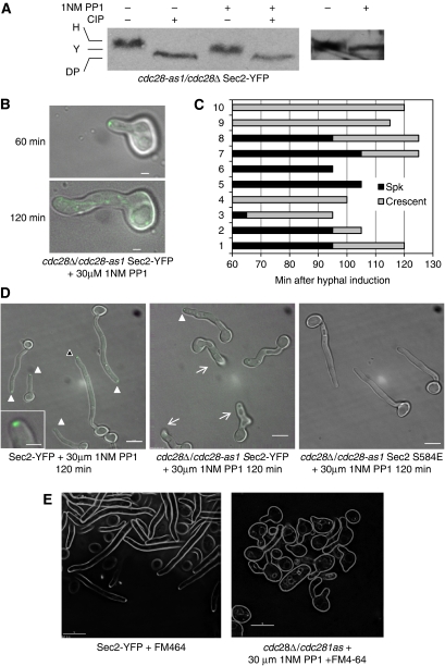
Sec2 phosphorylation and localization is Cdc28 dependent. (A) When Cdc28-1as is inhibited, Sec2-YFP shows the yeast, but not the hyphal pattern of phosphorylation upon hyphal induction. Stationary phase cells were size fractionated by elutriation and induced to form hyphae. Samples were collected after 180 min. (B–D) Sec2 localization and hyphal formation is inhibited by 1NM-PP1 in cells expressing only Cdc28-1as. Stationary phase cdc28-1as/cdc28Δ SEC2-YFP; CDC28/CDC28 SEC2-YFP or cdc28Δ/cdc28-as1 SEC2 S584E cells were induced to form hyphae in the presence of 30 μM 1NM-PP1 on the surface of agarose in concavity microscope slides and incubated in parallel at 38°C on a microscope stage heated by a temperature hood. (B) The same representative cdc28-1as/cdc28Δ SEC2-YFP cell imaged after 60 and 120 min as indicated. (C) 60 min after induction, 10 cdc28-1as/cdc28Δ SEC2-YFP cells were selected, their morphology and pattern of Sec2-YFP localization recorded at intervals. The duration of the Spitzenkörper or crescent pattern localization pattern is graphically represented by black bars or grey bars, respectively. Cessation of any apical localization is shown by the termination of the grey bars. (D) After 120 min, representative fields of cells of each genotype were recorded. Solid arrows show the presence of a Spitzenkörper, an example, highlighted by the black arrowhead, is enlarged in inset of the left panel. Barbed arrows in the middle panel show cells, which cannot be recorded in a single focal plane because the hypha has grown into the agarose substratum. In these cells, care was taken to examine them by using an extended focal range to verify the absence of any apical Sec2-YFP localization. (E) Inhibition of Cdc28 1as by 1NM PP1 prevents Spitzenkörper formation. Spitzenkörper was visualized by uptake of FM4-64, 90 min after hyphal induction. Scale bars: panel B, 1 μm; panel D, E, 5 μm; inset 1 μm.

