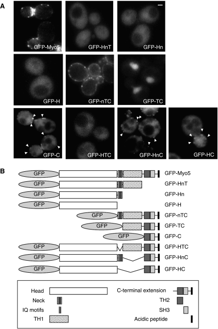
Figure 1.
Subcellular localization of GFP-Myo5 constructs. (A) Fluorescence micrographs of live myo5Δ cells (SCMIG275), expressing the indicated GFP fusion proteins represented in (B) expressed from centromeric plasmids under the control of the MYO5 promoter. Cells were grown to mid-log phase at 25°C and observed by conventional fluorescence microscopy. Arrowheads indicate cortical patches. Bar=1 μm.

