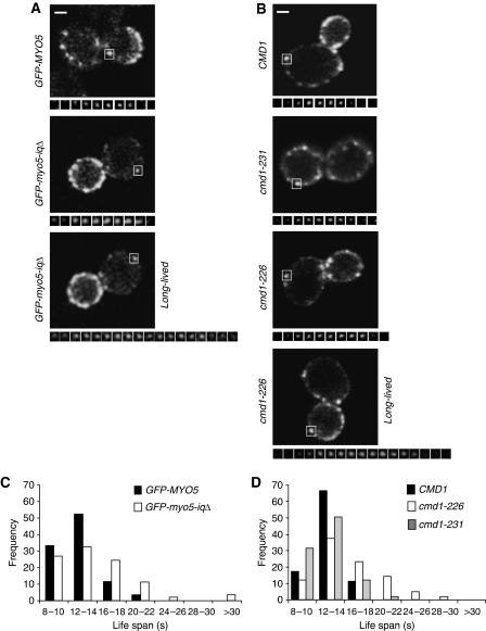
Figure 4.
Cmd1 dissociation from Myo5 expands its lifespan at cortical patches. (A, B) Fluorescence micrographs of consecutive frames from time-lapse movies of cortical patches (lower panels) of GFP-Myo5 or GFP-Myo5-iqΔ, expressed in a myo5Δ strain (Y06549) (A) or GFP-Myo5 expressed in a cmd1Δ myo5Δ strain, expressing the wt CMD1 (SCMIG1057) or the cmd1-231 (SCMIG1059) or cmd1-226 (SCMIG1058) mutants (B). The upper panels show a single frame of the imaged cells. The white square frames the cortical patch under analysis. Frames were recorded every 2 s. Constructs were expressed on centromeric plasmids under the MYO5 promoter. (C, D) Frequency distribution for the lifespan of the 80 GFP-Myo5 cortical patches analysed for the strains described in (A, B), respectively. Bar=1 μm.

