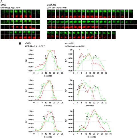
Figure 7.
Cmd1 dissociation from Myo5 triggers actin assembly at endocytic sites. (A) Fluorescence micrographs of consecutive frames from double-colour time-lapse movies of three representative cortical patches in cmd1Δ myo5Δ strains with RFP-tagged ABP1, expressing GFP-Myo5 from a centromeric plasmid under the MYO5 promoter and either the wt CMD1 (SCMIG1063) or the cmd1-226 mutant (SCMIG1064). Frames were recorded every 2 s. GFP-Myo5 patches with lifespans longer than 16 s were analysed for the cmd1-226 strain. (B) Graphs demonstrating the relative fluorescence intensity (RFI) plotted against time for the Abp1-RFP (red) and GFP-Myo5 (green) double-colour time-lapse movies shown in panel (A). ta and td indicate the arrival and departure times of GFP-Myo5 relative to ABP1-RFP, respectively.

