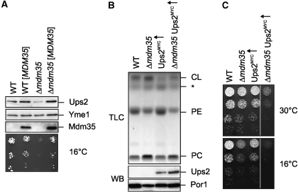
Figure 3.
Mdm35 controls the accumulation of Ups2 in mitochondria. (A) Steady-state levels of Ups2MYC in cells containing different amounts of Mdm35. Wild-type (WT) and Δmdm35 cells expressing genomically tagged Ups2MYC were transformed with either YCplac111∷ADH (PY127, PY129) or YCplac111∷ADH encoding MDM35 ([MDM35]) (PY128, PY130). Cells were grown to mid-log phase in selective synthetic glucose media. Proteins were extracted from mid-log phase cells by alkaline lysis, analysed by SDS–PAGE and immunoblotting using Yme1-, MYC- and affinity-purified Mdm35-specific antibodies (upper panel). Five-fold serial dilutions of these cells were spotted on selective, glucose-containing plates and incubated at 16°C (lower panel). (B, C) Ups2 overexpression does not restore normal PE levels and cell growth in the absence of Mdm35. (B) TLC analysis of mitochondrial lipids in wild-type (WT, CG214), Δmdm35 (CG524), Ups2MYC↑ (CG626) and Δmdm35 Ups2MYC↑ (PY64) cells (upper panel). The asterisk (*) indicates an unidentified lipid species. Mitochondria purified by sucrose-gradient centrifugation were used for determination of the phospholipid profile and were analysed by SDS–PAGE and immunoblotting using porin- (Por1) and MYC-specific antibodies (lower panel). (C) Five-fold serial dilutions of mid-log phase cultures of the cells were spotted on YP plates containing galactose and incubated at 16 or 30°C.

