Figure 5.
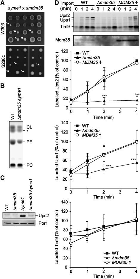
Mdm35 controls cell growth and the lipid composition of mitochondrial membranes in an Yme1-dependent manner. (A) Genetic interaction of MDM35 and YME1. Diploid Δmdm35Δyme1 cells that were obtained by mating of Δmdm35 and Δyme1 cells (CG324 × VIA5, CG560 × CG113) were sporulated and meiotic spores were grown on glucose-containing medium. White arrows indicate double-mutant progenies in the W303 strain background or inviable double-mutant spores in the strain S288c. (B) Reduced PE and CL levels in Δmdm35Δyme1 mitochondria. Phospholipid profiles of wild-type (WT, CG1) and Δyme1Δmdm35 (CW414) mitochondria were analysed by TLC. The asterisk (*) indicates an unidentified lipid species. (C) Ups2 accumulates in Δmdm35Δyme1 cells. Mitochondria were isolated from wild-type (WT, CG1), Δmdm35 (CG323), Δyme1 (VIA4), and Δmdm35Δyme1 (CW414) cells grown on galactose-containing media and analysed by Tris/Tricine gradient SDS–PAGE and immunoblotting using porin (Por1)- and affinity-purified Ups2-specific antibodies. (D) Accumulation of Ups2 in mitochondria upon import in vitro depends on Mdm35. Radiolabelled Ups1, Ups2 and, for control, Tim9, were imported into wild-type (WT, CG1), Δmdm35 (CG323) and Mdm35-overexpressing (CW343) mitochondria for the indicated time. The analysis of the samples by Tris/Tricine gradient SDS–PAGE and autoradiography is shown in the upper panels. A quantification of imported Ups1, Ups2 and Tim9 is shown in the lower panels. Protein imported into wild-type (WT) mitochondria after 4 min was set to 100%. Data represent±standard deviation of six independent experiments. ***P<0.0005.

