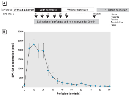
Figure 3.
The kinetics of BPA-GA in the maternal–placental–fetal unit after uterine perfusion. (A) Time schedule of uterine perfusion with 2 μM BPA-GA; an additional perfusion without BPA-GA was performed for 70 min after a 20-min perfusion with BPA-GA. (B) Time course of concentration of BPA-GA in the collected perfusate; after 70 min, BPA-GA was barely detected in the perfusate (n = 4).

