Figure 5.

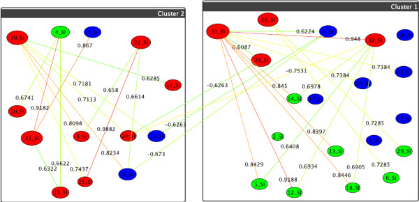
Graph-clustering correlation network for gangliosides showing the difference between U87+DI312/24hr+SN38/24hr and U87+p53/24hr+SN38/24hr. Correlation network resulting from graph-clustering data analysis for a correlation threshold > 0.6 for gangliosides showing the difference between the first two treatments (i.e., edge weights = cor(T1) - cor(T2)). The clustering for T1 (boxes) automatically divides into small-value (cluster 2) and high-value lipids (cluster 1), see Table 1. T2's clustering (colors) distinguishes further: high values (green nodes), intermed. values (blue nodes), and very small values (red nodes). Thus, red nodes in the right hand box show the strongest decrease when switching to T2. In particular, v31 = GD1 (d18:1/24:0) and v32 = GD1 (d18:1/24:1) show the highest change. in T1 they react similarly as several green nodes (e.g., v5 = GM3 (d18:1/16:1)), which they do not do for T2, as evident from the many heavy-difference edges incident to nodes v32 and v31. Also significant are v30 = GD1 (d18:1/23:1), v22 = GM1b (d18:1/23:0) and v21 = GM1b (d18:1/22:0), which, by their very small values in T2 set themselves apart from many others (heavy edges).
