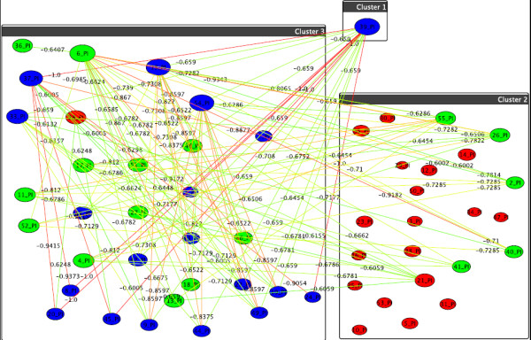
Figure 9.

Graph-clustering correlation network for phosphatidylinositol showing the difference between U87+DI312/24hr+SN38/24hr and U87+p53/24hr+SN38/24hr. Correlation network resulting from graph-clustering data analysis for a correlation threshold > 0.6 for PIs showing the difference between the first two treatments. Despite of the visual clutter, we here retain threshold for the sake of comparability to the above figures. Quite obviously, T1 and T2 heavily differ, with almost 7% of all pairwise differences above 0.6. Red nodes are mostly small, which indicates little discrimination between T1 and T2 for these nodes, which in the raw data correspond to lipids with high values. Quite noticeable, v39 = (38:6)+O shows the highest increase in T2 simply because its value is 0 in T1 but 100 in T2 while v6 = (34:2)+O, v15 = (36:3)+O, v37 = (38:5)+3O and v54 = (40:6)+O are also significant.
