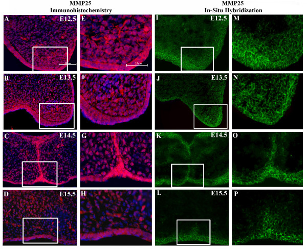
Figure 2.
Localization of matrix metalloproteinase-25 (MMP-25) protein and mRNA expression in the developing mouse palate. (A-H) Immunofluorescent images of MMP-25 protein expression (red) colocalized with Hoechst nuclear staining (blue). (I-P) In situ hybridization of MMP-25 mRNA expression (green). Expression of MMP-25 appears stronger in the epithelium of the palate shelves than in the underlying mesenchyme. (E-H) Enhanced views of highlighted areas from A-D. (M,P) Enhanced views of highlighted areas from I-L. For A-D and I-L, scale bar indicates 50 μm. For E-H and M-P, scale bar indicates 25 μm.

