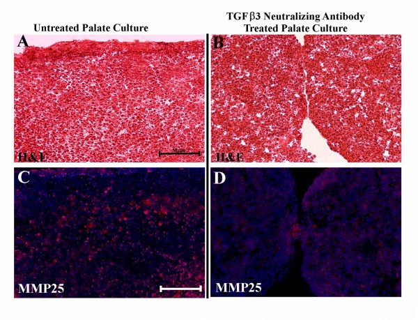
Figure 6.
(A, C) Embryonic day (E) 13.0 in vitro cultured control palate after 72 h, (B,D) E13 in vitro cultured palate (treated with TGF-β3-neutralizing antibody, 10 μg/mL) after 72 h. (A,B) Hematoxylin and eosin (H&E) stained sections. (C,D) Immunohistochemistry of MMP-25 (red) colocalized with Hoechst nuclear staining (blue). TGF-β3-neutralizing antibody-treated culture inhibited palatal fusion (B) and showed weaker MMP-25 protein expression (D). Scale bar indicates 50 μm.

