Figure 7.
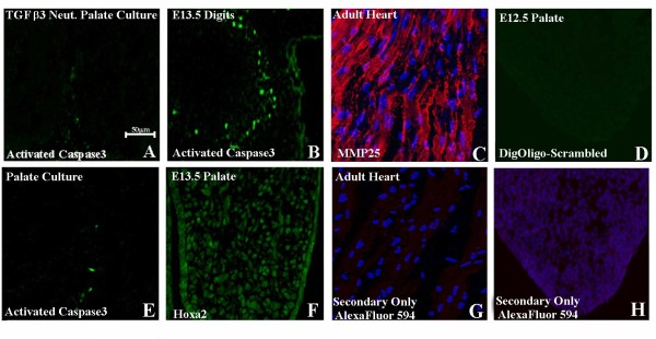
(A,B,E) Immunohistochemical staining for activated caspase3. Little or no activated caspase 3 could be identified in control palatal cultures (E) or in TGF-β3-neutralizing antibody-treated palatal culture (A). (B) Section of E13.5 digits exhibiting caspase3 activity as positive control. (C) Immunohistochemistry (IHC) section of adult mouse heart exhibiting MMP-25 expression (red) as positive control colocalized with Hoechst nuclear staining (blue). (G) IHC section of adult mouse heart with only the secondary antibody as negative control. (D) ISH of E12.5 palatal section with scrambled Dig-labeled oligo probe as negative control. (F) IHC of E13.5 palate showing Hoxa2 expression as positive control [4]. (H) IHC of E12.5 palatal culture with only the secondary antibody colocalized with Hoechst nuclear staining (blue) as negative control. Scale bar indicates 50 μm.

