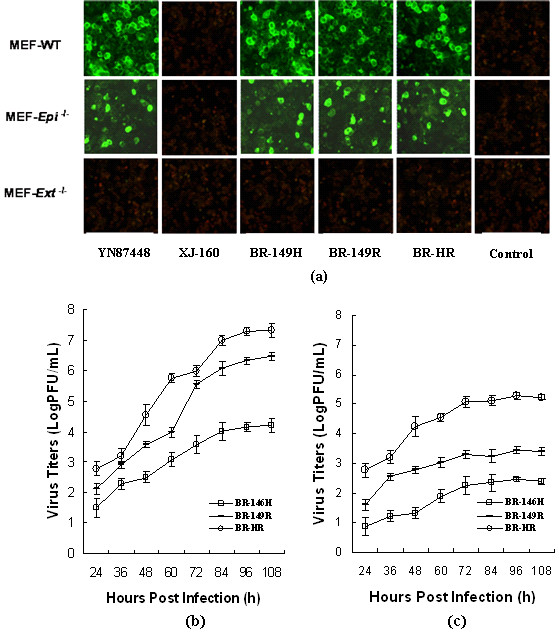
Figure 1.

Infectivity of recombinant viruses on MEF cell. (a) Comparison of viral infections to MEF cells. MEF cells were grown on cover slips to 80% confluent and infected with YN87448 and XJ-160 as well as the mutants for 48 hours. Antiserum against YN87448 (used in YN87448) or XJ-160 (used in XJ-160 panel and each of recombinant virus) diluted1:100 were applied for IFA as previously described [16]. Non-infected MEF cells were used as control; Monolayer of MEF-wt (b) and MEF-Epi-/-cells (c) were infected with recombinant or parental viruses at a MOI of 0.01, the medium (1 ml) was removed at the indicated time points and evaluated for virus titer by plaque assay. Each point represents the mean ± SD of three wells.
